

ADVERTISEMENT
Filtered By: Lifestyle
Lifestyle
Cancer scare? Not all breast lumps are created equal
Women Talk
Discovering a lump in one’s breast is scary for most women. The fear of it being cancerous is always there, especially if we have learned of female relatives or friends dying of this dreaded malignancy. Or some of us may have read Angelina Jolie undergoing a double mastectomy to lower her risks of getting breast cancer.
 To understand where these lumps come from, it is best to know its anatomy first.
Externally, what we see are (1) the nipples, (2) the dark area around it called the areola, and of course, the skin.
Internally, the breast is composed of the following:
(3) lactiferous alveoli or sacs that produce milk during lactation (after all, suckling of the young for survival is the foremost role of the breasts)
(4) lactiferous ducts that bring the milk from the sacs to the nipples
(5) sweat glands
(6) oil glands
(7) fat tissue, which gives the breast its form and size, while supporting the ducts
(8) the pectoralis, or chest muscles underneath.
During the premenstrual period, the breast may feel engorged and painful. This is due to normal hormonal fluctuations during the menstrual cycle.
Lumps and bumps
Breast masses can be solid, cystic (containing fluid), or both. Whatever their composition, they can be either benign or malignant. To ease your worries, it is best to know the different types of lumps.
The breast is mostly fat tissue, so any masses from fat could give rise to a lump, which is a benign growth.
If the breast hits a hard object, this could cause a rupture of the small blood vessels and can cause hematoma, a small lump filled with blood.
Sometimes, bacteria can enter the breast tissues through the nipples, or tears in the skin due to scratching or wounds, causing infection. This could lead to abscess formation, and signs of inflammation such as redness and swelling may be observed.
The sweat and oil glands too, could swell if their ducts are blocked. Again, if bacterial infection occurs, a woman may experience swelling of the breast.
From the milk glands, fibroadenomas or intraductal papillomas—benign growths due to excessive connective tissues—can form. The milk ducts themselves could also dilate – this is called ductal ectasia – producing a benign lump.
Sometimes a woman can continue to produce milk long after her delivery, a condition known as galactorrhea. If lactation persists, a doctor may request for a cranial CT SCAN to rule out a tumor in the pineal gland, which is a small gland in the brain.
Many times, patients consult me because of “breast pain” without lumps. The worry about the “Big C” comes up, but upon examination, it usually turns out that the pain is not on the breast itself, but on the ribs below the chest muscles. These patients, usually in their mid-forties, have inflamed cartilages of the ribs or costochondritis, not unlike arthritis of the bones. The pain is exacerbated when raising the arm on the affected side, but although the discomfort is sometimes extreme, it responds to analgesics.
What to do when you have lumps
The best thing to do when one finds a breast lump is to consult a doctor, who will do a thorough breast examination with the patient lying down, and sitting up. This is not only to verify the presence of the mass, but also to check for the size, consistency, location of the mass, and presence or absence of pain. Furthermore, the nipples will also be examined to see if there are discharges.
If you feel uncomfortable being examined even by a female doctor, by all means bring along a close friend, or a female relative to accompany you. The doctor will appreciate your openness on this matter.
Tests and doctor's diagnosis
If breast lumps are found, the doctor will advise you on what to do. Some will request for further mammography to look for calcifications, or hardening of the lumps. Breast ultrasound may also be undertaken to see if the lump is cystic or solid, and to localize the mass, especially if the breast is dense and large. In some cases, an MRI (Magnetic Resonance Imaging) may be done.
Some doctors will advise you to see a surgeon for further work-ups. The surgeon could advise you to simply observe the lump. But if the mass is cystic, an FNAB (Fine Needle Aspiration Biopsy) or an excision biopsy (removing the whole lump) may be done. All liquids and tissues will be sent to the pathologist, who will process the material and examine them under the microscope to see if there is evidence of malignancy; this is the only way to diagnose breast cancer.
Breast cancer among Pinays
One of the leading causes of death in females in the Philippines is still breast cancer. Age is considered the highest risk factor: it usually occurs in women who are 40 years old and above, and is manifested in many different ways.
A lump is usually felt, either by the female herself, or her partner. There is no way, however, to distinguish a benign from a malignant growth by simply feeling the lump. Bloody discharge from the nipple, “orange peel” layer (areas of the skin are retracted inwards because of the malignancy), and inflammation of the surrounding skin are all late signs of breast cancer.
A positive family history is a warning bell for many women, which brings us to early detection.
Doing breast self-examination (BSE)
All women are advised to examine their breasts regularly. The best time is after menses, when the breasts are soft. For those already in their menopause, select a specific and consistent day of the month for this self-examination.
I usually ask my patients to do their breast exam under the shower with a soapy hand, so that it could easily glide around the breast. Raise one arm and examine the breast of that side with the other hand. Imagine the breast as the face of a clock, and begin at 12 o’clock (uppermost, center). Press gently on the breast tissue, and slide the hand along the skin and move to 1 o’clock, then to 2, until all the areas are covered. We are feeling for lumps, and for pain. Press the nipple gently and note for any discharges. Do the same on the other breast.
Because breast cancer rarely occurs in the younger age group (below 40 years old), small lumps in these women are usually just observed. For those 40 and above, however, a yearly mammography is a good screening procedure; in the US, this has been cut down to once every two years.
Regular screening, plus the BSE, are good methods for early detection and will help any woman maintain peace of mind. – With illustration by Analyn Perez/YA, GMA News
Dr. Alice M. Sun-Cua is a writer and practicing obstetrician-gynecologist at the San Juan de Dios Hospital in Pasay City
Discovering a lump in one’s breast is scary for most women. The fear of it being cancerous is always there, especially if we have learned of female relatives or friends dying of this dreaded malignancy. Or some of us may have read Angelina Jolie undergoing a double mastectomy to lower her risks of getting breast cancer.
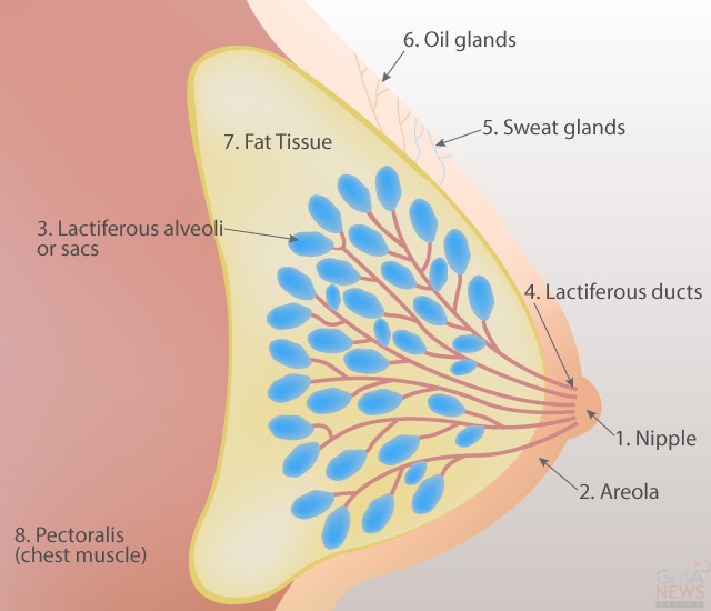
To understand where these lumps come from, it is best to know its anatomy first.Externally, what we see are (1) the nipples, (2) the dark area around it called the areola, and of course, the skin.
Internally, the breast is composed of the following:
(3) lactiferous alveoli or sacs that produce milk during lactation (after all, suckling of the young for survival is the foremost role of the breasts)
(4) lactiferous ducts that bring the milk from the sacs to the nipples
(5) sweat glands
(6) oil glands
(7) fat tissue, which gives the breast its form and size, while supporting the ducts
(8) the pectoralis, or chest muscles underneath.
During the premenstrual period, the breast may feel engorged and painful. This is due to normal hormonal fluctuations during the menstrual cycle.
Lumps and bumps
Breast masses can be solid, cystic (containing fluid), or both. Whatever their composition, they can be either benign or malignant. To ease your worries, it is best to know the different types of lumps.
The breast is mostly fat tissue, so any masses from fat could give rise to a lump, which is a benign growth.
If the breast hits a hard object, this could cause a rupture of the small blood vessels and can cause hematoma, a small lump filled with blood.
Sometimes, bacteria can enter the breast tissues through the nipples, or tears in the skin due to scratching or wounds, causing infection. This could lead to abscess formation, and signs of inflammation such as redness and swelling may be observed.
The sweat and oil glands too, could swell if their ducts are blocked. Again, if bacterial infection occurs, a woman may experience swelling of the breast.
From the milk glands, fibroadenomas or intraductal papillomas—benign growths due to excessive connective tissues—can form. The milk ducts themselves could also dilate – this is called ductal ectasia – producing a benign lump.
Sometimes a woman can continue to produce milk long after her delivery, a condition known as galactorrhea. If lactation persists, a doctor may request for a cranial CT SCAN to rule out a tumor in the pineal gland, which is a small gland in the brain.
Many times, patients consult me because of “breast pain” without lumps. The worry about the “Big C” comes up, but upon examination, it usually turns out that the pain is not on the breast itself, but on the ribs below the chest muscles. These patients, usually in their mid-forties, have inflamed cartilages of the ribs or costochondritis, not unlike arthritis of the bones. The pain is exacerbated when raising the arm on the affected side, but although the discomfort is sometimes extreme, it responds to analgesics.
What to do when you have lumps
The best thing to do when one finds a breast lump is to consult a doctor, who will do a thorough breast examination with the patient lying down, and sitting up. This is not only to verify the presence of the mass, but also to check for the size, consistency, location of the mass, and presence or absence of pain. Furthermore, the nipples will also be examined to see if there are discharges.
If you feel uncomfortable being examined even by a female doctor, by all means bring along a close friend, or a female relative to accompany you. The doctor will appreciate your openness on this matter.
Tests and doctor's diagnosis
If breast lumps are found, the doctor will advise you on what to do. Some will request for further mammography to look for calcifications, or hardening of the lumps. Breast ultrasound may also be undertaken to see if the lump is cystic or solid, and to localize the mass, especially if the breast is dense and large. In some cases, an MRI (Magnetic Resonance Imaging) may be done.
Some doctors will advise you to see a surgeon for further work-ups. The surgeon could advise you to simply observe the lump. But if the mass is cystic, an FNAB (Fine Needle Aspiration Biopsy) or an excision biopsy (removing the whole lump) may be done. All liquids and tissues will be sent to the pathologist, who will process the material and examine them under the microscope to see if there is evidence of malignancy; this is the only way to diagnose breast cancer.
Breast cancer among Pinays
One of the leading causes of death in females in the Philippines is still breast cancer. Age is considered the highest risk factor: it usually occurs in women who are 40 years old and above, and is manifested in many different ways.
A lump is usually felt, either by the female herself, or her partner. There is no way, however, to distinguish a benign from a malignant growth by simply feeling the lump. Bloody discharge from the nipple, “orange peel” layer (areas of the skin are retracted inwards because of the malignancy), and inflammation of the surrounding skin are all late signs of breast cancer.
A positive family history is a warning bell for many women, which brings us to early detection.
Doing breast self-examination (BSE)
All women are advised to examine their breasts regularly. The best time is after menses, when the breasts are soft. For those already in their menopause, select a specific and consistent day of the month for this self-examination.
I usually ask my patients to do their breast exam under the shower with a soapy hand, so that it could easily glide around the breast. Raise one arm and examine the breast of that side with the other hand. Imagine the breast as the face of a clock, and begin at 12 o’clock (uppermost, center). Press gently on the breast tissue, and slide the hand along the skin and move to 1 o’clock, then to 2, until all the areas are covered. We are feeling for lumps, and for pain. Press the nipple gently and note for any discharges. Do the same on the other breast.
Because breast cancer rarely occurs in the younger age group (below 40 years old), small lumps in these women are usually just observed. For those 40 and above, however, a yearly mammography is a good screening procedure; in the US, this has been cut down to once every two years.
Regular screening, plus the BSE, are good methods for early detection and will help any woman maintain peace of mind. – With illustration by Analyn Perez/YA, GMA News
Dr. Alice M. Sun-Cua is a writer and practicing obstetrician-gynecologist at the San Juan de Dios Hospital in Pasay City
